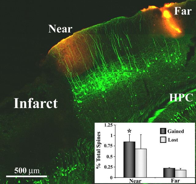
Figure 5.
Dendritic spine formation is elevated specifically within peri-infarct areas. Representative example of a fixed brain section from a YFP-transgenic mouse (sagittal plane, 2 weeks after stroke) where spine turnover was assessed near and far from the infarct border (sites demarcated with DiI, red-orange color). Inset, Graph showing increased levels of spine formation in regions proximal, but not distal to the infarct. *p < 0.05. HPC, Hippocampus.

