
Figure 1.
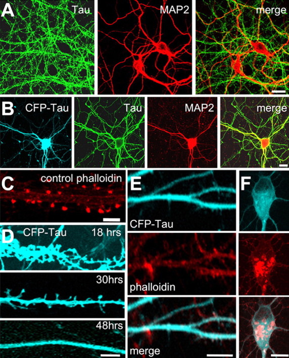
Tau distribution and changes in dendritic spine morphology after expression of tau. A, Untransfected hippocampal neurons. K9JA, an antibody for total Tau is localized only in axons, whereas MAP2 visualized by MAP2 clone HM2 antibody (Sigma) only localizes in dendrites. B, Expression of CFP-tau in hippocampal neurons leads to missorting into dendrites. CFP-tau appears in the soma and dendrites and colocalizes with MAP2 (HM2 antibody). C, Phalloidin fluorescence illustrates the concentration of F-actin in spine heads. D, After tau expression, CFP-tau appears along the dendrites and also in dendritic spines, followed by their disappearance (shown at 18, 30, and 48 h after transfection). E, F, Transfection by CFP-Tau for 3 d causes loss of spines and accumulation of F-actin in the dendritic shaft (E) and cell body (F) (staining by CFP-tau, phalloidin, and merge). Scale bars, 10 μm.