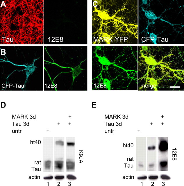
Figure 3.
Phosphorylation of tau in transfected neurons. Neuronal cultures at 25 div were transfected with tau for 2 d and then analyzed by immunofluorescence and Western blotting. A, Control cells (top) stained for total tau (antibody K9JA) and with antibody 12E8, which is sensitive to phosphorylated KXGS motifs in tau repeat domain. No antibody signal is detected for 12E8. B, After transfection with CFP-tau, cell bodies and all neurites show tau and react with 12E8 antibody. C, Double transfection with tau plus MARK2 reveal both proteins throughout the neurons and a strong 12E8 reaction. D, Western blots show reaction with the pan-tau antibody K9JA (transfected human htau40, top band; endogenous rat tau, bottom bands). E, Using the same membrane, the 12E8 reaction reveals phosphorylation at the KXGS motifs after transfection with tau, and especially with tau plus MARK2. Bottom bands are actin loading controls. Scale bar: (in C) A–C, 10 μm.

