
Figure 4.
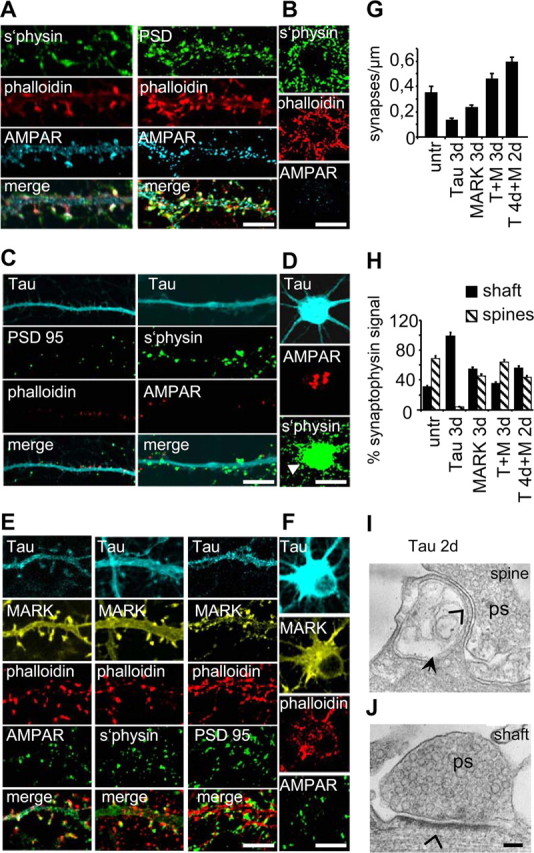
Immunofluorescence of presynaptic and postsynaptic proteins in transfected neurons. A Control cultures 25 div stained for AMPAR, PSD95, synaptophysin, and F-actin (phalloidin). B, No abnormal accumulation of markers seen in cell bodies of control cells. C, Tau-transfected neurons show strong reduction in AMPAR, PSD95, and synaptophysin after 3 d. D, A robust accumulation (arrowhead) of AMPAR and synaptophysin is seen in the cell bodies. E, F, In neurons transfected with tau plus MARK2, staining of all markers is similar to control in dendrites and cell bodies. G, Synapse densities determined from phalloidin (spine, postsynaptic) and synaptophysin positive puncta after 3 d of transfection. Tau transfected neurons show less synapses than doubly transfected neurons. H, Classification of synaptophysin-positive puncta reveal less shaft-synapses than spine-synapses in control cells. Tau transfected cells display only shaft synapses and tau plus MARK2 transfected cultures are similar to controls. I, Ultrathin section of a remnant of a spine 2 d after tau transfection. No PSD is detectable here nor in adjacent sections (open arrowhead) and the spine apparatus appears dilated (filled arrow). ps, Presynapse with vesicles. J, However, shaft synapses appear functional with visible PSD (open arrowhead). Scale bars: A–F, 10 μm; I, J, 150 nm. Error bars indicate SE.