Figure 5.
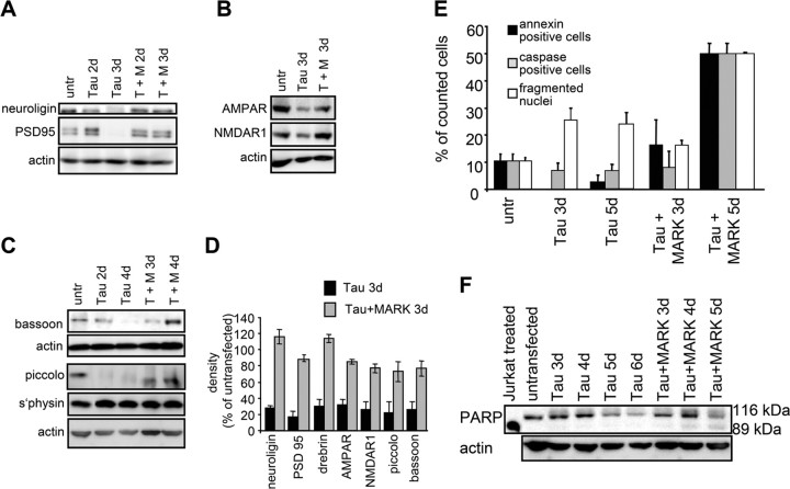
Presynaptic and postsynaptic proteins are reduced by tau expression but preserved by transfection of tau plus MARK2. A, Western blots showing that postsynaptic proteins neuroligin and PSD95 are reduced in tau-expressing cultures and that loss is prevented by double transfection of tau plus MARK2. The PSD95 antibody (MA1–045) detects a ∼95 kDa protein and a slightly larger species from rat brain extracts. Actin is shown as a control. B, C, Postsynaptic receptors NMDAR1, AMPAR, and the presynaptic proteins bassoon and piccolo are reduced in tau-expressing cultures during a time course of 2–3 d and rescued with double transfection of tau plus MARK2. Synaptophysin shows no overall change after tau transfection but accumulates in the cell body (Fig. 4D). D, Densitometric analysis of the Western blots for synaptic proteins (neuroligin, PSD95, drebrin, AMPAR, NMDAR1, piccolo, bassoon), all normalized to β-actin. The first bracket shows the reduction of protein levels after tau expression, compared with untransfected controls (n = number of culture preparations). The second set of parentheses shows the rescue in tau plus MARK2 transfected cultures: neuroligin (27 ± 3%; n = 10; *p < 0.001) (116 ± 23%; n = 7; *p < 0.05), PSD95 (17 ± 7%; n = 4; *p < 0.001) (88 ± 4%; n = 3; *p < 0.03), drebrin (30 ± 9%; n = 4; *p < 0.001) (115 ± 7%; n = 2; *p < 0.02), AMPAR (32 ± 8%; n = 5; *p < 0.001) (84 ± 8%; n = 5; *p < 0.005), NMDAR1 (26 ± 9%; n = 4; *p < 0.001) (77 ± 9%; n = 3; p > 0.05), piccolo (22 ± 13%; n = 2; *p < 0.001) (73 ± 18%; n = 2; p > 0.05), bassoon (26 ± 10%; n = 3; *p < 0.001) (77 ± 16%; n = 3; p > 0.05). E, F, Apoptotic markers are not significantly increased in tau-transfected neurons. E, Control cultures (25 div) stained for the markers annexin, active caspase, and DAPI. An increase of fragmented nuclei in tau-transfected neurons after 3 and 5 d of transfection is seen but no increase in annexin or active caspase. Doubly transfected neurons (tau plus MARK2) display no increase in these markers after 3 d, but a strong increase in staining after 5 d of transfection. F, PARP is downregulated in tau transfected neurons, but not degraded. In contrast, in doubly transfected neurons (tau plus MARK2), both the intact 116 kDa form and 85 kDa fragment of PARP are seen after 4 d of transfection. The control shows a cell extract of Jurkat cells induced to undergo Fas-mediated apoptosis by treatment with anti-human Fas mAb, clone DX2 (BD Biosciences, Franklin Lakes, NJ). Error bars indicate SE.

