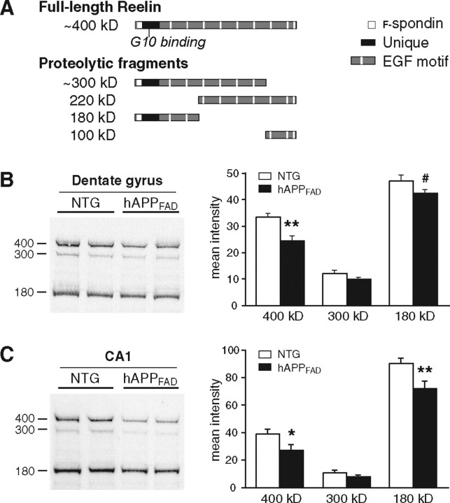
Figure 4.
Reelin levels are decreased in the hippocampus of hAPPFAD mice. A, Schematic illustrating full-length Reelin and the fragments produced by extracellular cleavage. Monoclonal antibody G10, used for both immunostaining and Western blot analyses, binds to Reelin where indicated. B, C, Western blot analysis of protein lysates from dentate gyrus (B) or CA1 (C) demonstrates a significant reduction in Reelin levels in hAPPFAD mice (n = 10 mice per genotype). Equal protein, determined by Bradford assay, was loaded in each lane. #p = 0.06; *p < 0.05; **p < 0.01 versus nontransgenic (NTG).

