Figure 5.
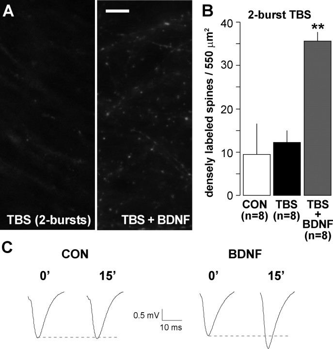
BDNF lowers the threshold for TBS-induced increases in spine F-actin. Hippocampal slices were treated with rhodamine–phalloidin for 20 min, after which baseline responses were collected and then TBS was applied. A, Photographs of rhodamine–phalloidin labeling show that two theta bursts did not increase numbers of densely labeled spines when applied alone (left) but did so when applied in the presence of 2 nm BDNF (right). Scale bar, 10 μm. B, Group data (mean ± SEM) confirm that slices receiving two theta bursts alone (TBS) had comparable numbers of F-actin dense spines as those receiving only baseline stimulation (CON; p > 0.3, two-tailed t test), whereas slices treated with two theta bursts in the presence of 2 nm BDNF (TBS + BDNF) had approximately threefold greater numbers of intensely labeled spines (**p < 0.01 vs TBS alone). C, Field EPSPs recorded immediately before (0′) and 15 min after (15′) delivery of two theta bursts to slices receiving aCSF (CON) or BDNF infusion (initiated 20 min before recordings).

