Figure 7.
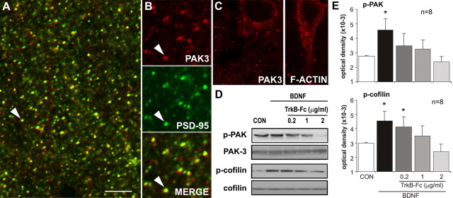
Exogenous BDNF promotes phosphorylation of actin regulatory proteins. A, High-power confocal micrograph shows anti-PSD-95 (green) and anti-PAK3 (red) double labeling in field CA1 (double-labeled elements appear yellow). Both markers labeled discrete puncta scattered throughout str. radiatum. Scale bar: A, 5 μm; B, 3 μm. B, Enlargements of the field including the arrow in A separated into red (PAK3), green (PSD-95), and overlaid (Merge) channels (A is a “merged” image) show that most, but not all, PAK3-immunoreactive puncta also contain PSD-95-IR. C, Micrographs show perinuclear localization of PAK3-IR (left) and phalloidin-labeled F-actin (right). D, E, Adult rat hippocampal slices were treated with BDNF (60 ng/ml; bath applied) for 30 min alone or with TrkB–Fc pretreatment. Representative Western blots (D) show treatment effects on p-PAK immunoreactivity (detects the conserved phosphorylation site on p-PAK isoforms 1, 2, and 3) with no evident changes in total PAK3. BDNF-induced increases in p-PAK were eliminated by TrkB–Fc in a dose-dependent manner. BDNF similarly increased, and TrkB–Fc similarly reduced, levels of p-cofilin without effects on total cofilin content. E, Quantification of Western blots assessing BDNF treatment effects on p-PAK (top) and p-cofilin (bottom) immunoreactivities, with and without TrkB–Fc cotreatment (group mean ± SEM band densities shown; n = 8 per group). In each case, BDNF-induced increases in the phosphoproteins (*p < 0.05 vs control, one-way ANOVA followed by Tukey's HSD test) were blocked by TrkB–Fc at concentrations from 0.2–2 μg/ml for p-PAK and 1–2 μg/ml for p-cofilin.

