
Figure 1.
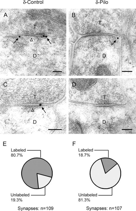
Electron micrographs of δ subunit immunogold labeling in the dentate molecular layer. A, C, Immunogold particles (arrows) indicate the location of the δ subunit on or near the plasma membrane of dendrites (D) that are in contact with axon terminals (T) in control mice. Gold particles are localized at perisynaptic sites, just outside or at the outer edge of symmetric synapses, and are found at either two ends (A) or one end (C) of the synaptic contact. Gold particles are not detected at central regions of the synapses (A, C, open arrowheads). B, D, In specimens from pilocarpine-treated mice, immunogold particles at many symmetric synapses had no immunogold labeling (D). When labeling was present, gold particles (arrow) were located at the outer edge of the synapse (B), as in controls. E, F, Quantitative analysis of symmetric synapses indicated that a high percentage of symmetric synapses were labeled for the δ subunit in control tissues (80.7%). In contrast, a low percentage of symmetric synapses were labeled for the δ subunit after pilocarpine treatment (18.7%). The δ subunit labeling was significantly less in pilocarpine-treated than in control mice (p < 0.005; χ2 test). Scale bars: A–D, 0.1 μm.