
Figure 3.
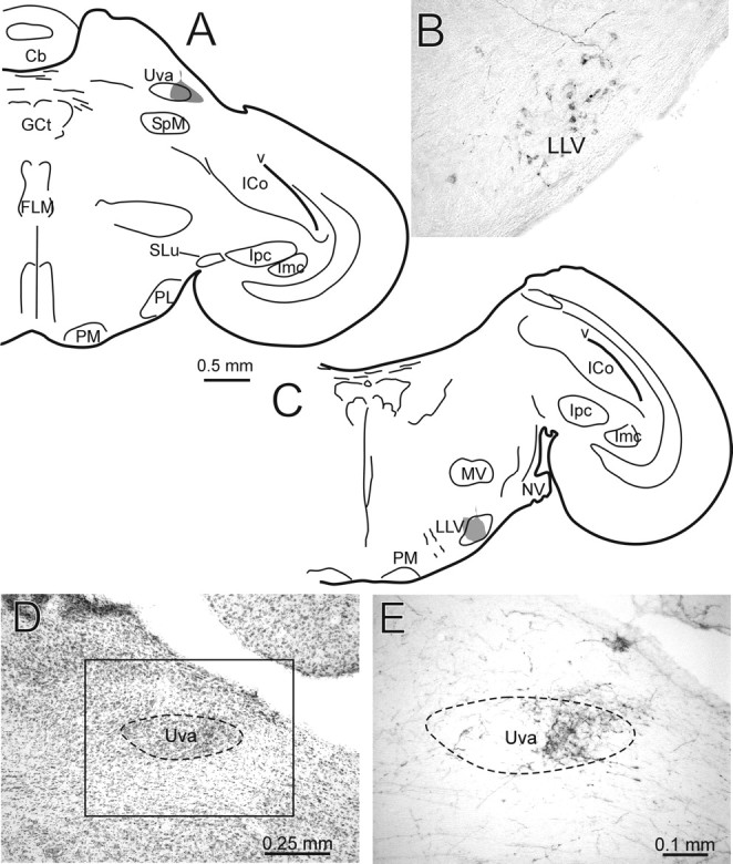
LLV projects to Uva. A, Schematic hemisection showing an injection of CTB (gray) centered on the lateral part of Uva. B, Photomicrograph of LLV neurons retrogradely labeled from the injection shown in A. Also obvious are several fibers labeled with BDA, an injection of which was made in a cochlear nucleus (nucleus angularis) in the same case. These fibers terminate within LLV. C, Schematic hemisection showing an injection of BDA (gray) in LLV. D, Nissl-counterstained section showing the grape-shaped nucleus Uva. Anterograde label resulting from the injection shown in C is present in the lateral part of Uva, which is shown in E at higher power in an adjacent non-counterstained section. E corresponds to the boxed area shown in D. Cb, Cerebellum; FLM, medial longitudinal fasciculus; GCt, central gray; ICo, intercollicular nucleus; Imc, magnocellular isthmic nucleus; Ipc, parvocellular isthmic nucleus; MV, rostral pole of trigeminal motor nucleus; NV, root of trigeminal nerve; PL, lateral pontine nucleus; PM, medial pontine nucleus; SLu, semilunar nucleus; SpM, medial spiriform nucleus; v, ventricle.