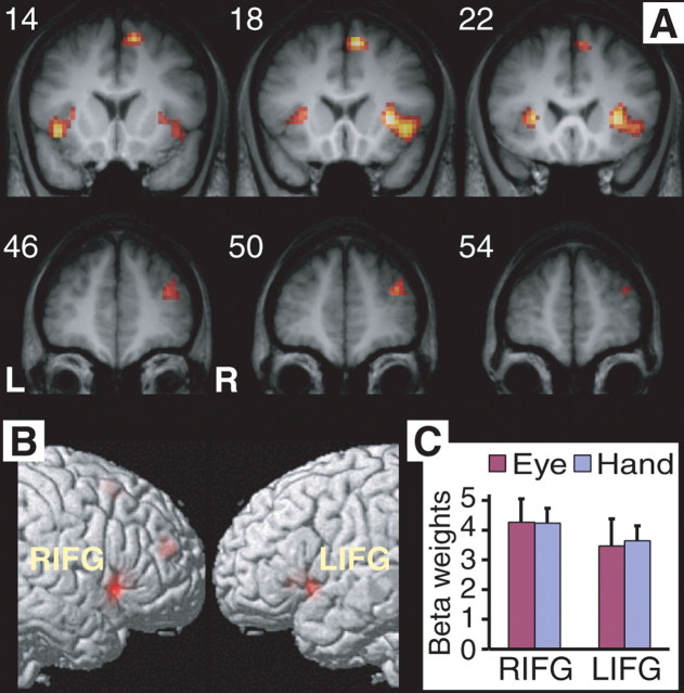
Figure 2.
Common activations in response inhibition. A, Suprathreshold activations were localized in the ventral IFG/insula in both manual and saccade stop–go contrast (p < 0.001, uncorrected). Common activations were also found in the MFG and medial SFG (see Table 1). Numbers indicate the y-coordinates in millimeters of the coronal slices. L, Left; R, right. B, Same results are shown on the lateral surface of the right and left hemispheres of the rendered MNI single-subject brain. C, Average beta values of the right IFG (RIFG) and left IFG (LIFG) clusters (in A) reveal similar level of activation in inhibiting hand and eye movements. Beta values are the parameter estimates of the SPM model (in arbitrary units). Error bars indicate the SEM.

