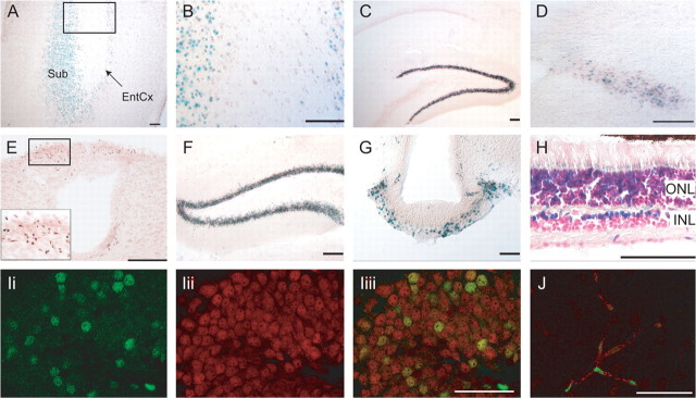
Figure 2.
β-Galactosidase expression in the CNS of postnatal and adult Cln3lacZ/+ mice. A, B, Subiculum and entorhinal cortex, P7. B is a high-power view of the box in A. C–E, Reporter expression at P21–P27. C, Hippocampus. D, Solitary tract. E, Area postrema; inset is high-power photomicrograph of boxed area. F–I, Reporter expression in adult (≥2 months old) brain. F, Dentate gyrus. G, Median eminence. H, Retina. I, A confocal image (0.37 μm slice) showing β-galactosidase (green) (Ii), NeuN (red) (Iii), and merged channels (Iiii) indicates Cln3 promoter activity in dentate gyrus granule neurons. J, A confocal image (0.37 μm slice) shows localization of β-galactosidase-positive nuclei (green) in CD31-positive vascular endothelial cells (red) in adult brain. Scale bars: A–H, 100 μm; I, J, 50 μm. Sub, Subiculum; EntCx, entorhinal cortex; ONL, outer nuclear layer; INL, inner nuclear layer.

