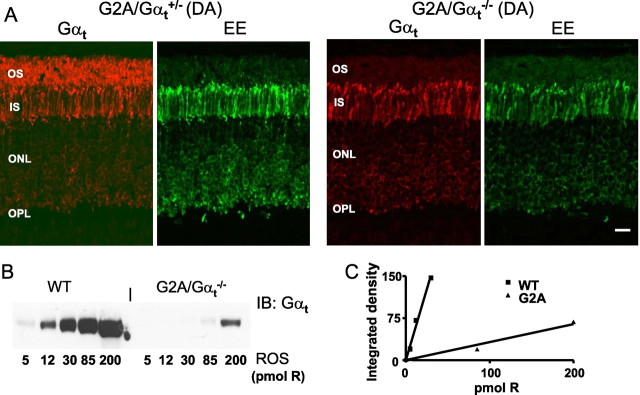
Figure 2.
A, Localization of total Gαt and mutant GαtG2A in dark-adapted (DA) mouse retinas. GαtG2A/Gαt+/− and GαtG2A/Gαt−/− retina sections were stained with rabbit anti-rod Gαt antibody K-20 or a monoclonal anti-EE antibody and visualized with goat anti-rabbit AlexaFluor 555 and goat anti-mouse AlexaFluor 488 secondary antibodies using a Zeiss LSM 510 confocal microscope. Scale bar, 20 μm. IS, Inner segment; ONL, outer nuclear layer; OPL, outer plexiform layer. B, Representative immunoblot (IB) analysis of relative levels of GαtG2A in GαtG2A/Gαt−/− ROS and native Gαt in WT ROS. C, Quantification of the relative protein levels from the immunoblot in B. The relative level of Gαt in WT ROS is ∼17-fold higher than the level of GαtG2A in transgenic ROSs (calculated as the ratio of the linear fit slopes). The average ratio of the slopes in three similar experiments is 18 ± 2 (mean ± SE).

