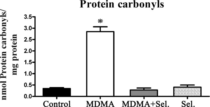
Figure 3.
MDMA induced carbonyl formation in whole-brain mitochondria and the protective effect of selegiline. Protein carbonyls were quantified by reaction with DNPH using a spectrophotometric assay for carbonyls. Animals were killed 14 d after exposure to: MDMA (4 × 10 mg/kg), selegiline (Sel.) (2 mg/kg) plus MDMA (4 × 10 mg/kg), saline (isovolumetric saline), or selegiline (2 mg/kg). Selegiline was administered 30 min before exposure to MDMA. Columns represent mean + SEM, expressed in nanomoles of protein carbonyls per milligram of total protein for each experimental group (n = 10 for control and MDMA; n = 6 for MDMA plus selegiline and selegiline). Animals exposed to MDMA presented significantly higher levels of carbonyl formation than all other groups (*p < 0.001, one-way ANOVA followed by a post hoc Scheffé's test).

