
Figure 3.
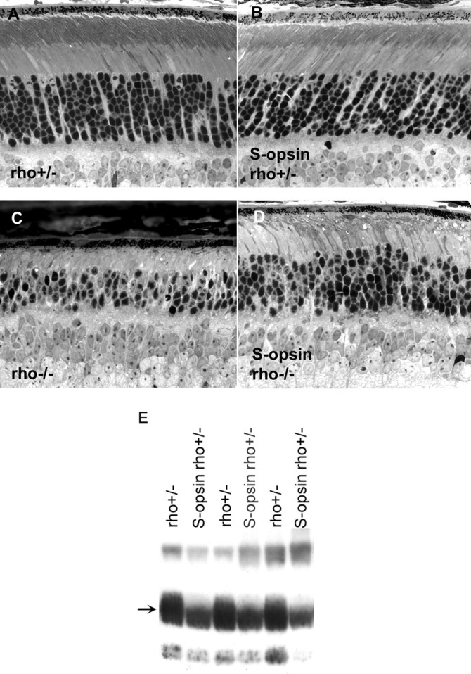
Analysis of retinal morphology and rhodopsin content in 2-month-old mice. A, Retinal morphology is largely normal in rho+/− mice. B, Retinal morphology is unchanged by the expression of S-opsin in this genetic background. C, The outer segment does not form in the absence of rhodopsin, and the number of rods decreases as a function of age. D, The presence of S-opsin delays retinal degeneration in rho−/− mice (compare C, D). E, Rhodopsin content is lowered when S-opsin is expressed; transgene-negative rho+/− littermates show higher rhodopsin content. Individual retinas were homogenized, and an equal fraction (1/3200) from each retina was loaded per lane. The proteins were blotted onto nitrocellulose and probed with 4D2, a monoclonal antibody against rhodopsin (arrow).