
Figure 1.
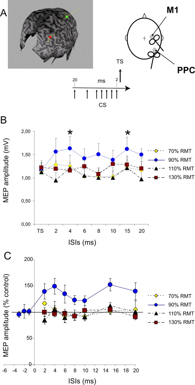
Effects of CS applied over the right PPC at different intensities on MEPs obtained by right M1 stimulation with subjects at rest. CS preceded TS applied over M1 by different ISIs ranging from 20 to 2 ms. The relative orientations of the coils are shown. In each case, the monophasic current pulse flows into the handle of the coil, inducing current in the opposite direction in the underlying cortex. In the MRI reconstruction, the most anterior point (green dot) is at the junction with the postcentral sulcus (hand area of the motor cortex), and the posterior point (red dot) lies over the angular gyrus. The yellow lines represent the ideal trajectories of the magnetic fields; these lines terminate at the presumed site of stimulation. A, Right PPC conditioning exerted potentiation over ipsilateral motor cortex. A single CS applied over the right PPC changed the amplitude of MEP obtained for ipsilateral M1 stimulation selectively when intensity of CS was set at 90% RMT with significant peaks obtained for ISIs of 4 and 15 ms. B, No significant change was observed for lower (70% RMT) or higher intensities of CS (110%, 130% RMT). Absolute values of MEPs in different conditions (mV) are shown in B, whereas data in C are normalized and expressed as percentages of control test conditions. Data obtained in experiment 3, when TS preceded by 1, 2, and 3 ms the CS set at 90% RMT intensity, are included in C. Errors bars indicate 1 SEM. Asterisks indicate a p value <0.05 at post hoc analysis.