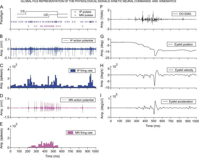
Figure 2.
Global file representation. A, A diagrammatic representation of the delay conditioning paradigm with indication of the time presentation for CS and US. Records illustrated in B–I were collected from the ninth conditioning session of two representative animals. The action potential pulses (IP pulses), marked with blue plus signs (A), correspond to the direct representation of the neuronal activity in the cerebellar interpositus nucleus (IP action potential, in B), and its respective instantaneous frequency (IP firing rate, in C). Action potential pulses (MN pulses) recorded from an orbicularis oculi motoneuron are indicated with magenta plus signs (A) and are the direct representation of the neuronal activity in the facial nucleus (MN action potential, in D) and its corresponding instantaneous frequency (MN firing rate, in E). The 24 kinetic parameters determined in this study (see Table 1) were collected from the firing activities of identified orbicularis oculi motoneurons (n = 110) and cerebellar interpositus (n = 174) neurons recorded across the successive habituation (n = 2), conditioning (n = 10), and extinction (n = 3) sessions from seven cats. F–I, These traces illustrate the electromyographic activity of the orbicularis oculi muscle (OO EMG, in F), the direct recording of the eyelid position by the magnetic field search-coil technique (G), and the estimated eyelid velocity (H) and acceleration (I) curves. The 36 kinematic parameters determined in this study (see Table 2) were collected from eyelid responses recorded across habituation, conditioning, and extinction sessions. For each of the physiological variables represented, the magnitude and the respective unit of measurement are indicated.

