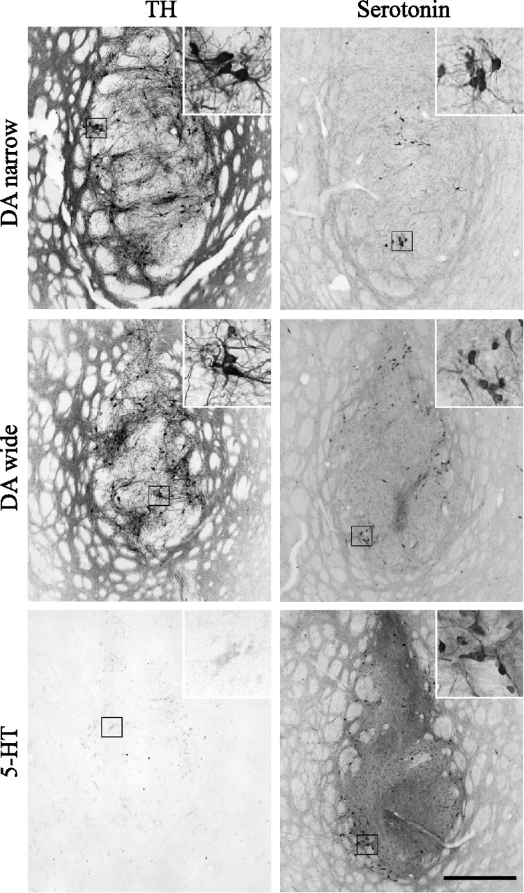
Figure 2.

Graft histology. Immunostaining for TH (A, C, E) and serotonin (B, D, F) revealed surviving cells in all grafted animals included in the study. The DA-narrow (A) and DA-wide (C) grafts contained large numbers of TH-positive neurons. In the 5-HT grafts group, in contrast, no or only scattered TH-positive neurons were detectable (E). The 5-HT grafts and the DA-wide grafts contained large numbers of serotonin-positive neurons, which were located both in the vicinity and the center of the graft core (D, F). The DA-narrow grafts, in contrast, showed significantly lower numbers of serotonin-positive neurons located mainly in the center of the graft core (B). High-power views showing single cells are illustrated as insets in each panel. Scale bar: (in F) A–F, low-power photomicrographs, 500 μm; insets, 125 μm.
