
Figure 3.
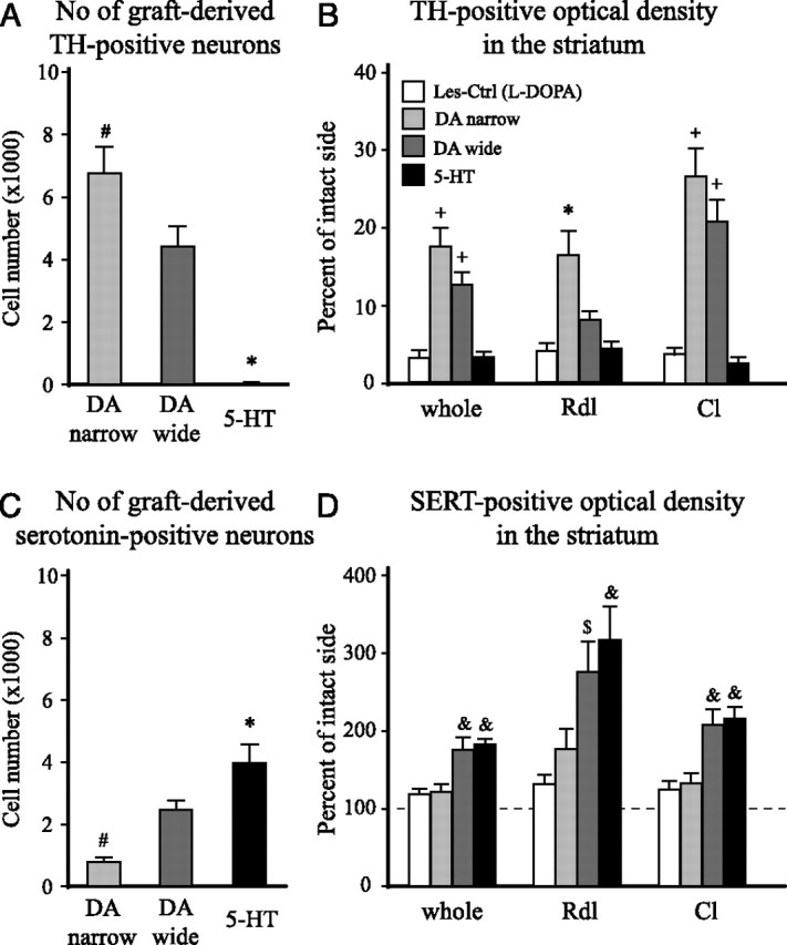
The total number of TH- and serotonin-positive cell numbers, and TH- and SERT-positive fiber density in the striatum. The cell numbers were determined using stereology (optical fractionator), and fiber innervation was assessed by optical density measurements. Fiber densities were measured in the whole striatum (excluding the graft core) and in two separate areas corresponding to the Rdl and Cl regions of the head of the striatum (indicated in Figs. 4A and 5A). The l-DOPA-treated lesion control group [Les-Ctrl (l-DOPA)] showed no TH- or serotonin-positive cells in the striatum at any level, and the TH-positive fiber innervation showed >95% depletion, in all regions, after the 6-OHDA lesion (B). The serotonin fiber density analysis, in addition, showed no significant hyperinnervations or hypoinnervations in any of the regions analyzed (118–127% of intact side) (D). The DA-narrow grafts contained significantly larger numbers of DA neurons than the DA-wide grafts (6732 ± 837 vs 4390 ± 640, respectively) (A). This was matched by an extensive TH-positive fiber innervation in the whole striatum and in the Cl region (DA narrow: whole, 18.2 ± 2.5%; Cl, 26.6 ± 3.6%; DA wide: whole, 13.0 ± 1.7%; Cl, 20.8 ± 2.8%, relative to the intact side) (B). The DA-narrow grafts also showed a significant reinnervation of the Rdl region (16.3 ± 3.1% of intact side), which was more pronounced than in the animals with DA-wide grafts (8.0 ± 1.0% of intact side). The 5-HT grafts displayed only scattered TH-positive neurons (7 ± 2), and no significant TH-positive fiber outgrowth in the striatum at any level (<5% of intact side in all regions) (A, B). The 5-HT graft group showed significantly larger numbers of serotonin-positive neurons in the graft (3865 ± 605) compared with both the DA-wide (2397 ± 286) and the DA-narrow grafts (767 ± 106), and the DA-narrow grafts showed significantly lower number of serotonin-positive neurons compared with the DA-wide grafted animals (C). SERT staining revealed a significant graft-derived serotonin innervation in the whole striatum in both the 5-HT (181.9 ± 14.3% of intact side) and DA wide (175.6 ± 15.4% of intact side) graft groups. The increase in SERT fiber density was evident in both the Cl region (5-HT, 208.9 ± 15.6; DA wide, 201.1 ± 20.3%, relative to the intact side) and, in particular, in the Rdl region (5-HT, 306.7 ± 42.4; DA wide, 266.7 ± 37.9% of intact side) (D). The DA-narrow grafts showed no significant graft-derived serotonin innervation in any region of the host striatum (whole, 121.4 ± 9.8%; Rdl, 170.9 ± 25.3%; Cl, 127.7 ± 13.3%, compared with intact side) (D). The dashed line in D represents the level of SERT-positive optical density on the contralateral intact side. *Different from all other groups. #Different from the DA-wide group. +Different from Les-Ctrl (l-DOPA) and 5-HT groups. &Different from Les-Ctrl (l-DOPA) and DA-narrow groups. $Different from Les-Ctrl (l-DOPA) group. A, C, One-way ANOVAs, F(2,29) = 31.49 and 14.39, respectively; p < 0.0001 for both comparisons. B, One-way ANOVAs, F(3,39) = 20.44, 10.99, and 26.66 for whole, Rdl, Cl, respectively; p < 0.0001 for all comparisons. D, One-way ANOVAs, F(3,39) = 8.04, 6.82, and 9.46 for whole, Rdl, Cl, respectively; p < 0.001 for all comparisons. All ANOVAs were followed by Tukey's HSD post hoc test. Error bars indicate SEM.