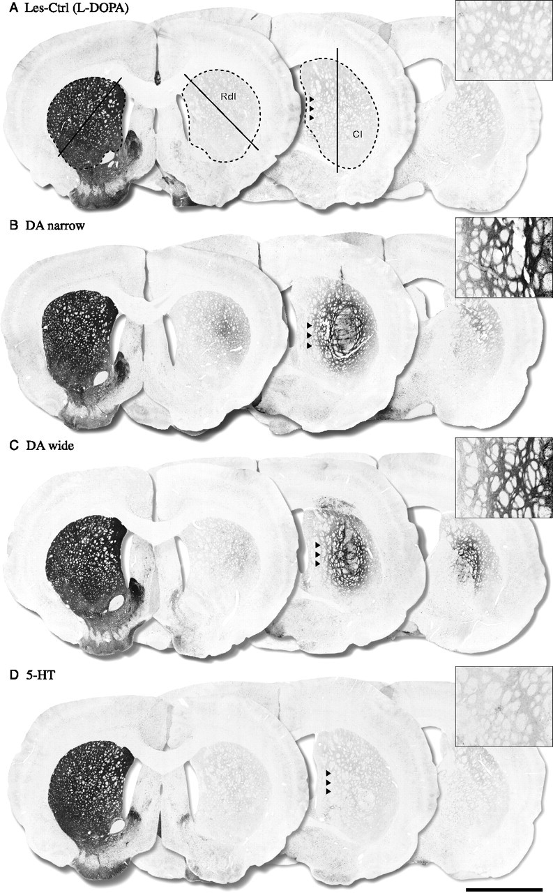
Figure 4.

TH-positive immunohistochemistry. The TH-positive fiber outgrowth was measured in the whole striatum and in two selected regions in the head of the striatum, the Rdl, and Cl as shown in A. The lesion control groups showed an almost complete loss of TH-positive fibers throughout the striatum with >90% denervation compared with the intact side (A) [Les-Ctrl (Drug-Naive) data not shown]. The groups receiving DA neuron-containing grafts (DA narrow and DA wide) showed significant TH-positive fiber innervations in the Cl region (40 and 30% of intact side, respectively) (B, C) and in the whole striatum (19 and 15% of intact side, respectively) (B, C). The DA-narrow grafts also displayed a significant increase in TH fiber density in the Rdl region (15% of intact side), which was not observed in the DA-wide group (8% of intact side) (B, C). The 5-HT graft group, in contrast, showed no TH-positive fiber outgrowth in the striatum in any region (<5% of intact side) compared with the control groups (D). The optical density values presented in brackets represent the animal from which the pictures are taken. Scale bar: (in D) A–D, 3 mm.
