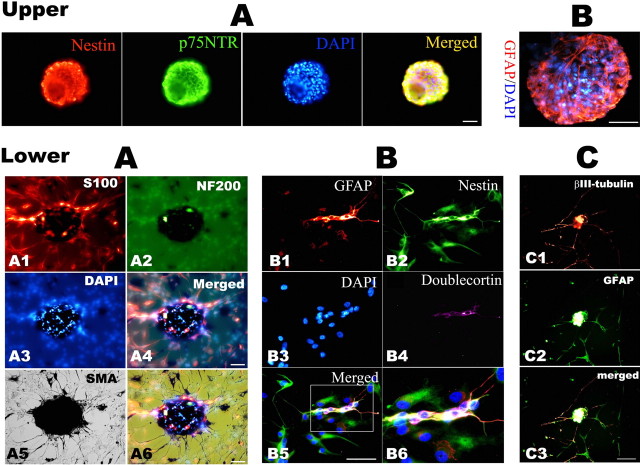
Figure 8.
Characterization by immunofluorescence of the secondary spheres derived from adult TG explants. Immunohistochemical characterization of neurospheres derived from adult rat TG explant cultures. Top, A, Representative photomicrographs show that spheres express Nestin and p75NTR; B, another sphere was stained for GFAP, and nuclei were counterstained by DAPI. Bottom, Immunocytochemistry staining for lineage markers on 2 week differentiation culture of hand-picked secondary spheres in DM. A, The same field image showing S100 (A1, red), NF200 (A2, green), and SMA (A5, black by DAB development) labeling; A4 is the merged image of A1–A3, and A6 is the overlay micrograph of A4 and A6. B, A representative staining of GFAP (B1, red), Nestin (B2, green), and DCX (B4, purple); B5 is the merged image of B1–B4, and B6 is the enlarged image of the white box inset in B5. C, Double staining of the differentiation culture with βIII-tubulin (C1, red) and GFAP (C2, green); C3 is the merged image of C1 and C2. Scale bar, 50 μm.

