
Figure 1.
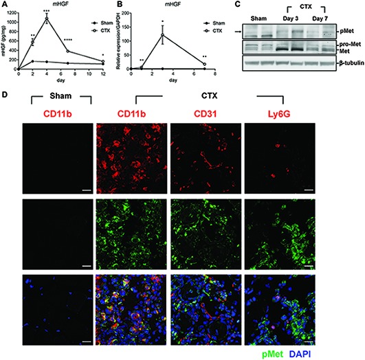
HGF/c-met signaling is activated during muscle regeneration. (A) Expression kinetics of HGF protein during muscle injury by CTX and regeneration. The muscle was isolated at 2, 4, 7, and 12 days after CTX injection, and total proteins were analyzed by ELISA to measure the protein levels of HGF. *p < 0.05, **p < 0.01, ***p < 0.001, ****p < 0.0001 versus sham-treated muscles (unpaired student’s t test), n = 4 per group. (B) Expression kinetics of the RNA levels of HGF during muscle injury and regeneration. RNA was prepared from TAs 1, 3, and 7 days after CTX injection followed by RT-qPCR, *p < 0.05, **p < 0.01 versus sham-treated muscles (unpaired student’s t test), n = 4 per group. The values were normalized to glyceraldehyde-3-phosphate dehydrogenase (GAPDH). (C) Expression kinetics of total and phosphorylated c-met proteins in CTX-injured TAs. Muscles were isolated at 3 and 7 days postinjury, and total proteins were prepared followed by western blot using specific antibodies to total or phosphorylated c-met. β-tubulin was used as a loading control. Each lane represents a sample from an individual mouse. Two representative mice are shown here. Two independent experiments were performed (with a total of four mice), and similar results were obtained. Arrow indicates the protein of interest in blots. (D) Identification of cell types expressing c-met. CTX-injured TA was isolated 3 days postinjury and subjected to immunofluorescence assay using antibodies to CD11b for macrophages, CD31 for endothelial cells, Ly6G for neutrophils (all red), and phosphorylated c-met (green). Nuclei were counterstained with DAPI (blue). n = 4 per group. Scale bars, 20 μm. All data are presented as mean ± SEM. See also Supplementary Figure S1.