
Figure 5.
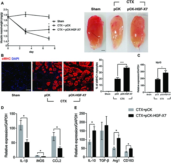
Effects of HGF overexpression by intramuscular injection of HGF expressing plasmid on muscle regeneration. pCK or pCK-HGF-X7 was i.m. injected 3 days prior to the CTX injection. TAs were prepared at appropriate times after injury. (A) Effect on TA weight. Representative TAs from 7 days postinjury are shown in the photos. *p < 0.05 versus CTX + pCK group (one-way ANOVA), n = 4 per group. Scale bar, 1 mm. (B) Effects on cross-sectional areas of regenerating fibers. CTX-injured TA was isolated 3 days postinjury and subjected to immunofluorescence assay using antibodies to eMHC (red) for regenerating myofibers. Percentage of eMHC+ myocytes was indicated in the graph. ***p < 0.001 (one-way ANOVA), n = 4 per group. Scale bar, 50 μm. (C–E) Three days after injury, the TA was isolated, and total RNAs were analyzed by RT-qPCR. (C) Effects on the expression of Myh3. *p < 0.05 (one-way ANOVA), n = 4 per group. (D) Effects on the RNA levels of M1 markers (IL-1β, iNOS, and CCL2). *p < 0.05 (one-way ANOVA), n = 4 per group. (E) Effects on RNA levels of M2 markers (IL-10, TGF-β, Arg1, and CD163). *p < 0.05 (one-way ANOVA), n = 4 per group. Values were normalized to GAPDH. All data are presented as mean ± SEM. See also Supplementary Figure S4.