
Figure 5.
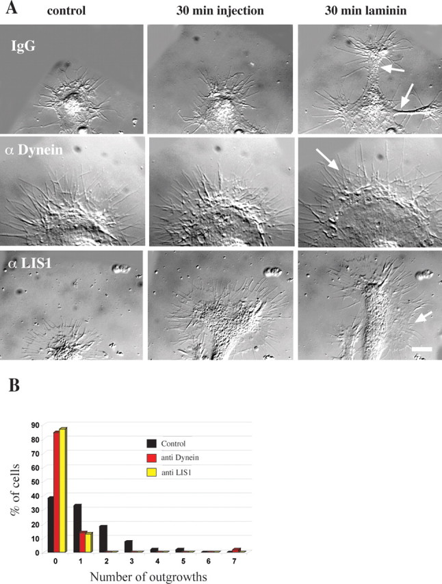
Effect of dynein IC and LIS1 antibody injection on growth cone morphology. A, Morphology of growth cones injected with antibody and then exposed to laminin. The growth cone of a neuron injected with control IgG shows a typical response. At 30 min after exposure to laminin two nascent axons have emerged (arrows). The growth cone of a cell injected with dynein IC antibody shows little change. After laminin treatment the growth cone has enlarged, but there are no outgrowths. The growth cone has a healthy peripheral actin network (arrow). A cell injected with polyclonal LIS1 antibody shows no outgrowths. After 30 min of exposure to laminin the growth cone has advanced a little, and lamellipodial regions are seen along the length of the newly formed neurite shaft (arrow). Scale bar, 10 μm. B, Quantitation of antibody injections. Chick DRG neurons were injected with monoclonal dynein IC antibody (n = 58), polyclonal LIS1 antibody (n = 31), or control mouse IgG (n = 55). Then the neurons were exposed to laminin for 30 min to induce neurite outgrowth. The number of laminin-induced neurites was reduced considerably in cells injected with function-blocking anti-dynein or anti-LIS1 antibody.