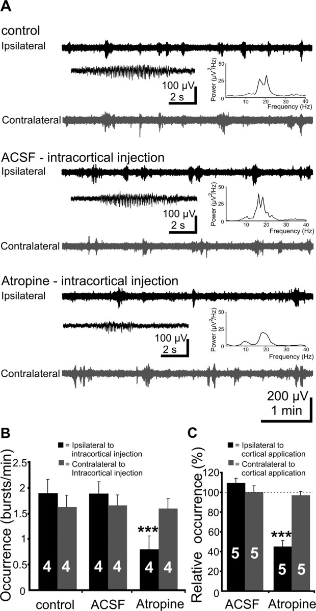
Figure 2.

Effects of mAChR blockade by intracortical injection or by application of atropine to the cortical surface on V1 spindle bursts. A, Extracellular field potential recordings from ipsilateral (black trace) and contralateral (gray trace) V1 of a P5 rat under control conditions, after intracortical injection of 20 nl of ACSF, and after intracortical injection of 20 nl of atropine (10 μg/g body weight, in ACSF). Inset, Typical spindle bursts displayed at an expanded time scale and averaged power spectrum of the field potential oscillations corresponding to the displayed traces. Note the reduced burst occurrence on the side of atropine injection. B, Bar diagram displaying the effects of intracortical atropine injection on the occurrence of bursts in the ipsilateral (black bars) and contralateral (gray bars) V1 of four P5–P6 pups. C, Bar diagram displaying the effects of atropine application to the V1 surface on the occurrence of bursts in the ipsilateral (black bars) and contralateral (gray bars) V1 of five P5–P6 pups. The occurrence of spindle bursts before cortical application was considered as 100%. The hemisphere of intracortical injection or cortical application was defined as ipsilateral.
