
Figure 2.
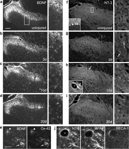
DRI induces BDNF and NT-3 expression in the dorsal horn. a, In the intact spinal cord, BDNF immunoreactivity was restricted to the terminals of primary afferent axons. Higher-power images from the same sections are shown on the right (boxes in lower-power images indicate their approximate origins). b–d, BDNF was upregulated in glial cells by 3 d after C7/8 DRI and remained elevated for at least 20 d. e, Ox-42 immunohistochemistry revealed BDNF expression by microglia in early (arrowhead) and intermediate (arrow) stages of reactivity. f–h, NT-3 immunoreactivity was intense in cerebellar Purkinje cells (inset), absent from the uninjured dorsal horn, but present in blood vessel-associated (asterisk) cellular processes as early as 3 d after DRI (g, h, arrows). h, i, DRI-induced NT-3 expression persisted for at least 20 d. j, NT-3 was expressed in glial fibrillary acidic protein (GFAP)-positive astrocytes around rat endothelial cell antigen 1 (RECA-1)-positive vasculature (a white-matter astrocyte from the degenerating dorsal columns is shown). Scale bars: a, 100 μm; e, i, 25 μm.