
Figure 3.
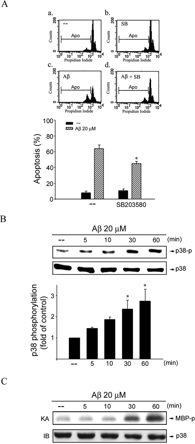
p38MAPK in Aβ-induced CEC apoptosis. A, CECs were pretreated with vehicle or 10 μm SB203580 (SB), a specific p38MAPK inhibitor, for 30 min before treatment with 20 μm Aβ for another 48 h. The percentage of apoptotic cells was then analyzed by flow cytometric analysis of PI-stained cells as described in Materials and Methods. Compiled results are shown at the bottom. Each column represents the mean ± SEM of at least three independent experiments. *p < 0.05 compared with the group treated with Aβ alone. B, CECs were treated with 20 μm Aβ for 0–60 min, and p38MAPK phosphorylation was determined by immunoblotting with anti-phospho-p38MAPK antibody. Equal loading in each lane is reflected by approximately similar intensities of p38MAPK at the bottom. *p < 0.05 compared with the control group. C, Cells were treated with 20 μm Aβ for 0–60 min, and p38MAPK activity was assessed using MBP as a substrate. After SDS-PAGE, γ-32P-labeled MBP was visualized by autoradiography. Typical bands representative of three independent experiments with similar results are shown. KA, Kinase assay; IB, immunoblotting; AP, apoptotic region.