
Figure 5.
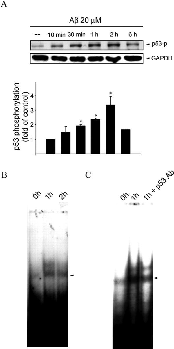
Aβ-induced p53 phosphorylation and increase in p53 binding activity in CECs. A, CECs were treated with 20 μm Aβ for indicated time periods. Cells were then harvested, and p53 phosphorylation at Ser15 was determined by immunoblotting with an anti-pSer15–p53 antibody. Equal loading in each lane is shown by the similar intensities of GAPDH. Compiled results are shown at the bottom. *p < 0.05 compared with the control group. B, CECs were incubated with 20 μm Aβ for 1 and 2 h. After incubation, the nuclear protein fraction was prepared for EMSA as described in Materials and Methods. C, An anti-p53 antibody (Ab) was included before EMSA to detect the specificity of p53 binding activity. Data shown are representative of three independent experiments with similar results.