
Figure 7.
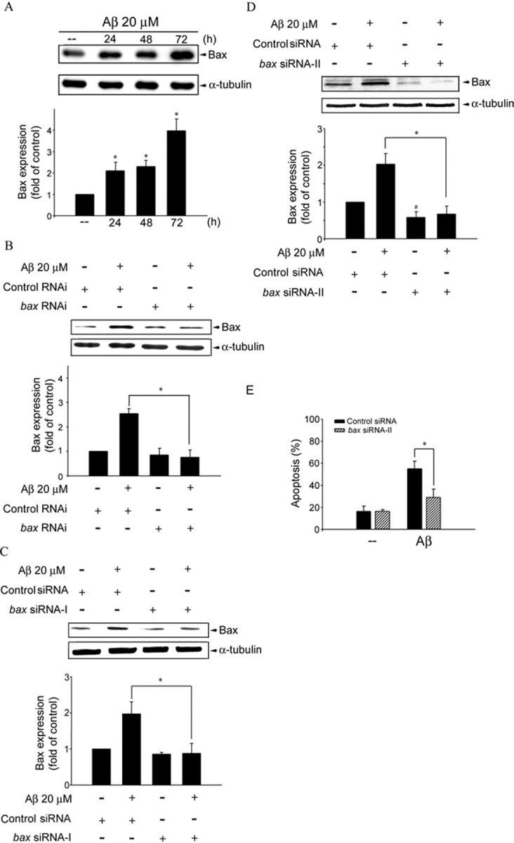
Bax expression in Aβ-induced CEC death. A, CECs were treated with 20 μm Aβ for 24–72 h and then harvested for assessing the extent of Bax expression by immunoblotting with an anti-Bax antibody. Compiled results are shown at the bottom. *p < 0.05 compared with the control group. B, CECs were transfected with control RNAi or bax RNAi for 24 h. After transfection, cells were treated with vehicle or 20 μm Aβ for another 48 h. The expression of Bax was then determined by immunoblotting with an anti-Bax antibody. Compiled results are shown at the bottom. *p < 0.05 compared with the control RNAi group in the presence of Aβ. C, CECs were transfected with control siRNA or bax siRNA-I for 24 h. After transfection, cells were treated with vehicle or 20 μm Aβ for another 48 h. The expression of Bax was then determined by immunoblotting with an anti-Bax antibody. Compiled results are shown at the bottom. *p < 0.05 compared with the control siRNA group in the presence of Aβ. D, CECs were transfected with control siRNA or bax siRNA-II for 24 h. After transfection, cells were treated with vehicle or 20 μm Aβ for another 48 h. The expression of Bax was then determined by immunoblotting with an anti-Bax antibody. Compiled results are shown at the bottom. *p < 0.05 compared with the control siRNA group in the presence of Aβ. #p < 0.05 compared with the control siRNA group. E, CECs were transiently transfected with control siRNA or bax siRNA-II for 24 h. After transfection, cells were treated with vehicle or 20 μm Aβ for another 48 h, and cell apoptosis was determined by flow cytometry. *p < 0.05 compared with the control siRNA group in the presence of Aβ.