
Figure 8.
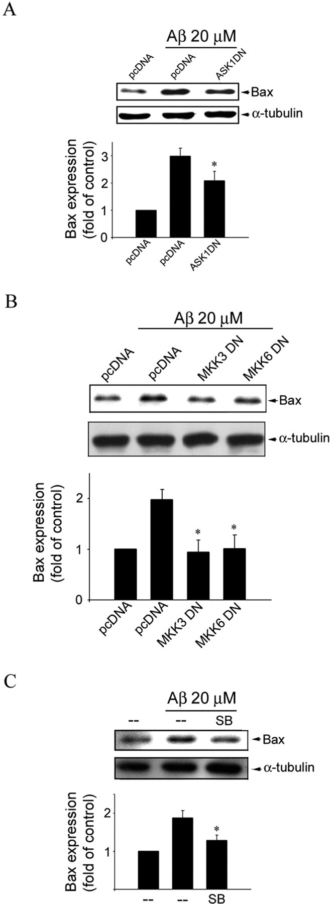
ASK1, MKK3, MKK6, and p38MAPK in Aβ-induced Bax expression in CECs. A, Cells were transiently transfected with pcDNA or ASK1 DN for 24 h. After transfection, cells were treated with vehicle or 20 μm Aβ for another 48 h. Cells were then harvested, and the extent of Bax expression was determined by immunoblotting with an anti-Bax antibody. Equal loading in each lane is shown by approximately similar intensities of the α-tubulin bands. Compiled results are shown at the bottom. *p < 0.05 compared with the pcDNA (mock transfection) group in the presence of Aβ. B, CECs were transiently transfected with pcDNA, MKK3 DN, or MKK6 DN for 24 h. After transfection, cells were treated with vehicle or 20 μm Aβ for another 48 h and then harvested for assessing the extent of Bax expression as described in A. Equal loading in each lane is shown by approximately similar intensities of the α-tubulin bands. Compiled results are shown at the bottom. *p < 0.05 compared with the pcDNA (mock transfection) group in the presence of Aβ. C, CECs were pretreated with vehicle or 10 μm SB203580 (SB), a specific p38MAPK inhibitor, for 30 min, followed by vehicle or 20 μm Aβ for another 48 h, and then harvested for assessing the extent of Bax expression as described in A. Equal loading in each lane is shown by approximately similar intensities of the α-tubulin bands. Compiled results are shown at the bottom. *p < 0.05 compared with the pcDNA (mock transfection) group in the presence of Aβ.