
Figure 8.
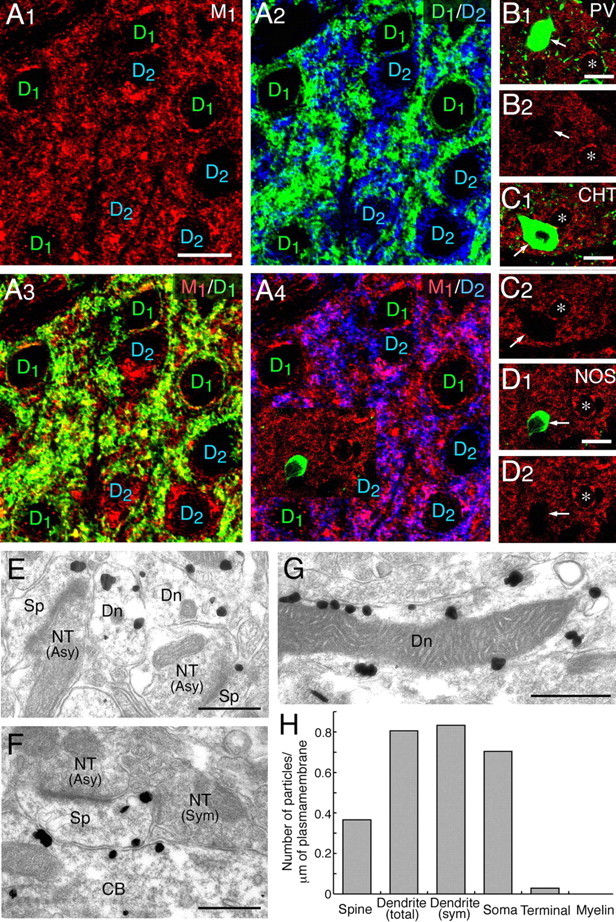
Muscarinic M1 receptor is preferentially expressed in the somatodendritic compartments of D1- and D2-positive MS neurons. A, Triple labeling for M1 (red), D1 (green), and D2 (blue) receptors. Note colabeling of M1 with D1 or D2 receptors in thin perikaryal rims and neuropils. Scale bar, 10 μm. B–D, Arrows indicate interneurons expressing PV (B), high-affinity CHT (C), or neuronal NOS (D). Note vacant M1 staining in these interneurons in contrast to abundant M1 expression in perikarya of putative MS neurons (asterisks). Scale bars, 10 μm. E–G, Silver-enhanced immunogold microscopy for M1 receptor. Note preferential M1 labeling in spines (Sp), dendritic shafts (Dn), and cell bodies (CB) of putative MS neurons, in contrast to negative labeling in nerve terminals (NT) forming asymmetrical (Asy) and symmetrical (Sym) synapses. Scale bars, 1 μm. H, The mean number of metal particles per 1 μm of the cell membrane on spines (measured length, 177.5 μm), total dendrites (173.9 μm), dendrites within 500 nm from the edge of symmetrical synapses (12.0 μm), soma (29.8 μm), nerve terminals (71.5 μm), and myelin sheath (33.4 μm).