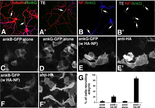
Figure 4.
NF and ankG are detergent resistant in the IS and preferentially bind to each other in HEK 293 cells. A, B, Live detergent extractions of neurons. A, Neurons were either fixed directly (A, B) or extracted live in Triton-X100 (A′, B′; TE) before fixation and then stained against MAP2 (blue), tubulin (red), and ankG (green) (A, A′), or against MAP2 (blue), NF (red), and ankG (green) (B, B′). AnkG and NF are retained at the IS (arrows). C–G, Ankyrin recruitment assays in HEK 293 cells. HEK 293 cells were transfected with ankB-GFP alone (C), with ankG-GFP alone (D), with ankG-GFP plus HA–NF (E, E′), or with ankB–GFP plus HA–NF (F, F′). Ankyrin distribution was visualized in the green channel (C–F), whereas HA–NF was detected with an anti-HA antibody (E′, F′). G, Quantification of three independent experiments at equivalent ankyrin expression levels. The percentage of cells showing ankyrin recruitment is plotted on the y-axis for various combinations of expressed proteins as indicated on the x-axis. Error bars show the SDs.

