
Figure 8.
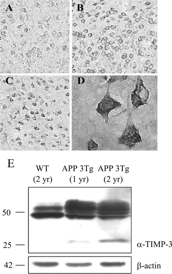
TIMP-3 immunoreactivity is increased in a mouse model of AD. Brain tissue from a mouse model of AD was immunostained for TIMP-3. Neuronal TIMP-3 expression increased with the age of these mice (A, 6 months; B, 12 months; C, 20 months; cortical neurons, original magnification, 20×). TIMP-3 immunostaining was perinuclear and punctate in cortical neurons. D, There was also a strong increase in TIMP-3 immunostaining in hippocampal neurons of the AD mouse model (63×). E, Brain tissue from wild-type (WT) or APP triple-transgenic (APP 3Tg) mice was extracted in RIPA buffer, and proteins were analyzed by Western blot with an anti-TIMP-3 antibody. Top, Both 25 and 50 kDa TIMP-3 species were increased in the AD mouse model compared with the wild-type mouse. Bottom, β-Actin was measured as a loading control.