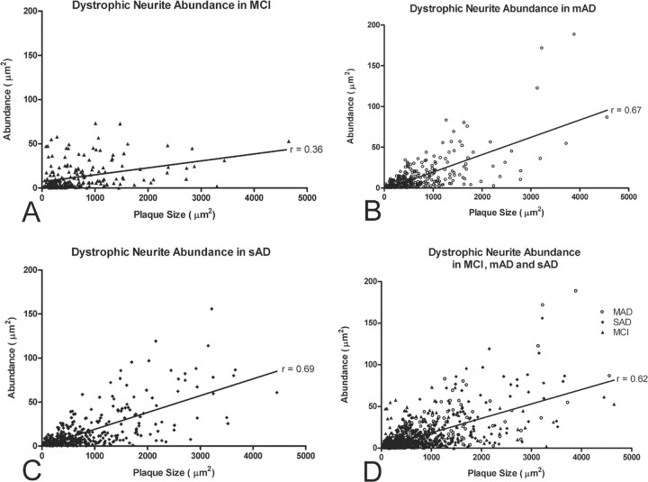
Figure 4.
Graphic depiction of the association between glutamatergic dystrophic neurite abundance and amyloid plaque size in the midfrontal gyrus of subjects with MCI (A), mAD (B), sAD (C), or all three subject groups combined (MCI, mAD, and sAD; D). Note the positive association between increasing plaque size and increasing glutamatergic dystrophic neurite abundance in all three groups. Note also that amyloid plaque size appears to increase with disease progression, as does the strength of the Pearson's correlation, thus suggesting that increasing levels of fibrillar Aβ correlate with increased glutamatergic terminal toxicity.

