
Figure 5.
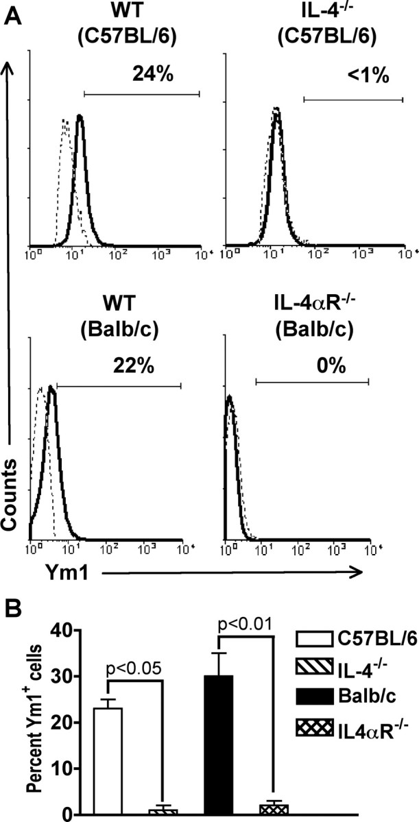
Ym1 protein expression in microglial cells is IL-4 dependent. Total mononuclear cells were isolated from the CNS of groups of four to five C57BL/6, IL-4−/−, BALB/c, and IL-4αR−/− mice and stained for expression of surface markers and Ym1. A, The solid line represents staining with anti-Ym1, and the dotted line represents background staining using rabbit serum. The horizontal bar indicates positive staining, and the number above the bar is the percentage positive. One representative experiment of three is shown. B, Mean ± SD of three separate experiments. There was a statistically significant decrease in the percentage of Ym1+ microglial cells in both IL-4- and IL-4αR-deficient mice when compared with wild-type control animals, as determined by the unpaired t test.