
Figure 7.
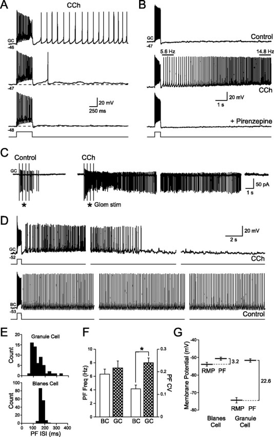
Persistent activity in granule cells after muscarinic receptor activation. A, Depolarizing granule cells slightly (from −48 to −46 mV) converts the step-evoked ADP response into a prolonged action potential discharge. Responses to 50 pA steps recorded in 2 μm CCh. B, Persistent firing evoked by 50 pA depolarizing steps in CCh is abolished by the M1 receptor antagonist pirenzepine (10 μm). Granule cell firing frequency was not constant and increased threefold during the response shown in the middle panel. C, Moderate intensity glomerular layer stimulation (4 × 100 μA; 400 ms interval) triggered spiking in a cell-attached granule cell recording. Bath application of CCh (2 μm) converted this transient response into a prolonged discharge that lasted >40 s. Gaps between consecutive sweeps shown were 20 s. D, Comparison of the persistent firing modes of granule cells (GC; top traces; recorded in 2 μm CCh; 80 pA step) and Blanes cells (BC; bottom traces; recorded under control conditions; 120 pA step). Both cells held at approximately the same membrane potential and stimulated with 500 ms depolarizing steps. Gaps between consecutive sweeps shown were 10 s for the GC and 20 s for the BC. The same voltage and time calibration for granule and Blanes cell recordings was used. E, Histograms of the interspike intervals in the response after the depolarizing steps (PF ISI) in the granule and Blanes cells shown in D. F, Summary of persistent firing frequency (PF Freq) and the coefficient of variation of the instantaneous firing frequency (PF CV) for granule cells in CCh and Blanes cells in control conditions. *p < 0.05. G, Summary of the differences between the RMP and persistent firing threshold (PF) for Blanes and granule cells.