Figure 3.
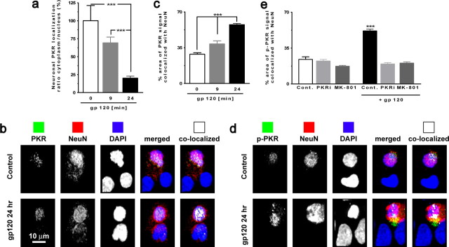
PKR and p-PKR translocation to nucleus in neurons exposed to gp120. a, PKR colocalization with MAP-2 (cytoplasmic) and DAPI (nuclear) illustrates the PKR translocation to nuclei in cultures exposed to gp120. ***p < 0.001 for n = 4 experiments. b, Images acquired using confocal microscopy of neurons (NeuN positive; red) exposed to gp120 for 24 h with PKR (green) and counterstained with DAPI. Colocalized PKR with NeuN is shown in white in the far right panels. c, PKR colocalization with NeuN revealed an increase of PKR in nuclei after exposure to gp120 for 24 h. ***p < 0.001 for n = 4 experiments. d, Images acquired using confocal microscopy of neurons (NeuN positive, red) exposed to gp120 for 24 h with p-PKR (green) and counterstained with DAPI. Colocalized p-PKR with NeuN is shown in white in the far right panels. e, Increased colocalization of p-PKR and NeuN in neurons exposed to gp120 after 24 h, which is abrogated in the presence of PKRi or MK-801. ***p < 0.001 for n = 4 experiments. Cont., Control. All values are mean ± SEM.

