Figure 5.
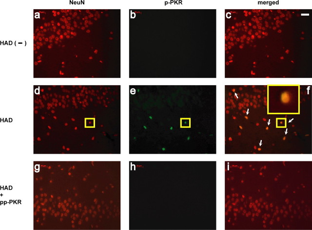
The activated form of PKRi localized in neuronal nuclei from autopsied brain sections of HAD patients. a–c, Hippocampal tissue from an HIV-positive but HAD-negative brain that had been double labeled with anti-NeuN (red) and anti-p-PKR (green) but no p-PKR labeling. d–f, Hippocampal tissue from an HAD patient manifested numerous NeuN-labeled cells colocalized with p-PKR (white arrows). In f, a magnification of the immunofluorescent image of one cell (small yellow box, d–f) is shown in the inset. g–i, Absence of p-PKR staining on HAD sections after incubation with pp-PKR. Results represent findings in brain sections from five HAD-positive individuals and five control patients including HIV infection but not HAD. Scale bar, 20 μm.

