
Figure 7.
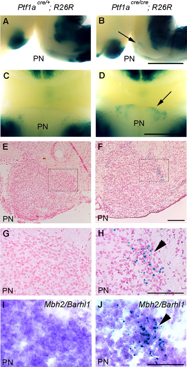
Fate change of Ptf1a lineage cells to MF neurons of the PN in Ptf1a null mutants at E18.5. Genotypes are indicated. A, B, Lateral views of whole-mount embryos stained with X-gal. C, D, Ventral views around the PN regions. Arrows in B and D indicate ectopically localized β-gal-positive cells in the mutant PN. E, F, Transverse sections of the PN regions stained with X-gal and nuclear fast red. G, H, High-magnification views of rectangular regions in E and F, respectively. I, J, Adjacent serial sections of G and H, respectively, which were stained with X-gal and subsequently subjected to in situ hybridization with Mbh2/Barhl1. Scale bars: A–D, 1 mm; E–J, 100 μm.