
Figure 7.
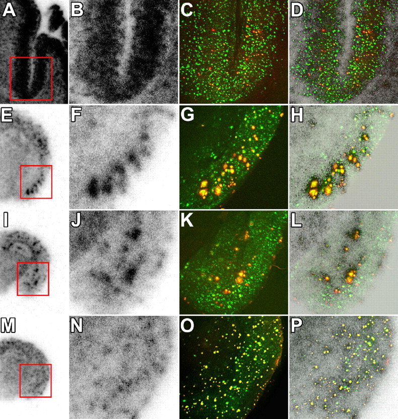
Association between in vitro radiolabeling of amyloid with [11C]PIB and abundance of Aβ40 and Aβ42. Brain sections from the AD patient (A–D), 23-month-old APP23 mouse (E–H), 23-month-old Tg2576 mouse (I–L), and 8-month-old PS-1/APP double-Tg mouse (M–P) were used for autoradiography with [11C]PIB (low-power and high-power autoradiograms are shown in the first and second columns from the left, respectively), and thereafter doubly immunostained for Aβ40 (red in the third column) and Aβ42 (green in the third column) with polyclonal anti-Aβ40 and monoclonal anti-Aβ42 antibodies. Colocalization of radiolabeling with Aβ40 and Aβ42 was assessed by merging autoradiograms with immunohistochemical data (shown in the fourth column). Similar double-immunofluorescence staining was obtained by using monoclonal anti-Aβ40 and polyclonal anti-Aβ42 antibodies (data not shown). Red squares indicate areas shown in magnified images.