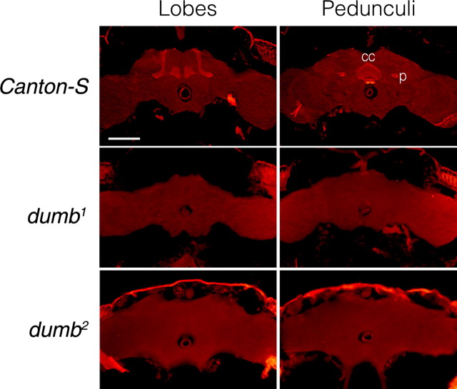
Figure 1.
dDA1 IR in the adult head sections of Canton-S, dumb1, and dumb2 flies. The frontal sections at the levels of the MB lobes (top) and pedunculi along with the central complex (bottom) are shown. dDA1 IR is visualized by red florescence. Canton-S has prominent dDA1 IR in the MB lobes and pedunculi as well as the central complex; however, no dDA1 IR is visible in those structures of dumb1 and dumb2. p, Pedunculus; cc, central complex. All images are at the same magnification. Scale bar, 100 μm.

