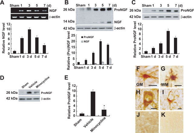
Figure 1.
Minocycline inhibits proNGF expression after SCI. A, B, NGF mRNA (A) and protein expression (B) were increased after SCI. B, C, Anti-mature-NGF antibody detected both mature (14 kDa) and proNGF (26 kDa) (B), and anti-proNGF antibody detected the high-molecular-weight band of 26 kDa (C). Note that the extent of induction of proNGF was greater than that observed with mature NGF (B). D, Minocycline treatment decreased the level of proNGF expression compared with that observed in vehicle-treated control at 5 d after injury. E, Quantitative analysis of Western blots shows that minocycline significantly inhibited proNGF expression when compared with that in vehicle control at 5 d after injury. Values are mean ± SD of three separate experiments. *p < 0.001. F–I, Immunocytochemical analysis shows that microglia in the white (WM) (F) and gray (GM) (G) matters, and neuron (H) and astrocyte (I) located near the lesion site expressed proNGF at 5 d after injury. J, K, Sham-operated (J) and negative control (K) were negative for proNGF. Double labeling using specific cell markers revealed that proNGF-positive cells shown in F–I were indeed microglia (F, G), neuron (H), and astrocyte (I), respectively (data not shown). Scale bars, 10 μm.

