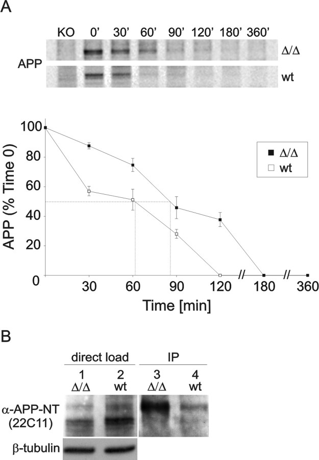
Figure 2.
Turnover of full-length APP and cell surface expression. A, Turnover of endogenous cell-bound APP. APPΔCT15-KI and wt MEFs were pulse-labeled with [35S]methionine/cysteine for 15 min and chased for the indicated time intervals. At time 0, APP consists predominantly of immature N-glycosylated species. Both the N-glycosylated and mature N- and O-glycosylated species are abundant at 30 min for both cell types. After a 60–90 min chase period the APP level is reduced dramatically in wt MEFs and below detection level at 120 min. In contrast, in APPΔCT15-KI MEFs the APP expression is reduced to only 40% after a 120 min chase period. Half-life was determined by quantitating the results as indicated by dotted lines (B). All values are presented as the means ± SEM. Differences were detected with two-tailed Student's t test, accepting p < 0.05 as significant. B, Biotinylation of surface APP. APPΔCT15-KI and wt MEFs were surface-biotinylated for 30 min. Cell lysate (15 μg/lane) was loaded directly onto a 10% Tris-tricine gel (lanes 1, 2) or was immunoprecipitated (350 μg/immunoprecipitate) by using NeutrAvidin-agarose beads (lanes 3, 4). APP was detected with the use of an N-terminal anti-APP antibody (22C11), revealing a considerable increase of surface APP in APPΔCT15-KI cells when compared with wt controls.

