
Figure 5.
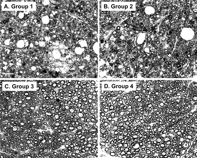
Bax deletion does not affect axon and myelin degeneration in Tg(F35) mice. Toluidine blue-stained, semithin, plastic sections of white matter from the cervical spinal cord were prepared from terminally ill, 90-d-old mice in group 1 (F35+/0 Prn-p0/0 Bax+/0) (A), group 2 (F35+/0 Prn-p0/0 Bax0/0) (B), group 3 (F35+/0 Prn-p+/0 Bax+/0) (C), or group 4 (Prn-p+/0 Bax+/0) (D) mice. Axonal loss, vacuolation, and myelin collapse are visible in groups 1 and 2 mice, whereas groups 3 and 4 mice show no abnormalities. Scale bar (in A) A–D, 20 μm.