
Figure 4.
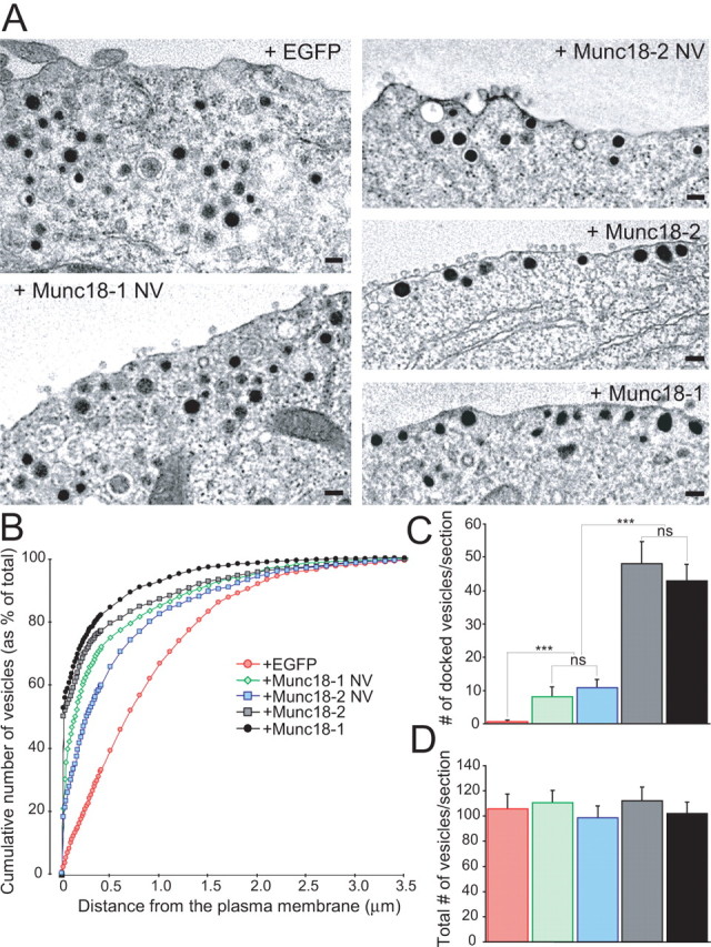
Stimulatory role of Munc18-1 binding to closed syntaxin1 in the docking step. A, The electron micrographs show the intracellular distribution of large dense-core vesicles close to the plasma membrane. munc18-1 −/− (null) cells expressed EGFP with or without variants of Munc18 proteins. Note that vesicles are not in close contact with the plasma membrane without Munc18s. Scale bars, 100 nm. B, Normalized cumulative distribution of vesicles as a function of distance from the plasma membrane. Data represent several cells/condition (see below). C, D, The number of docked vesicles (C) and the total number of vesicles (D). The data show that the ability of Munc18 to bind to syntaxin1 correlates with the level of rescue of vesicle docking. Data are mean ± SEM from the following number of cells (n) and animals (N): EGFP, n = 19, N = 4; Munc18-1 NV, n = 22, N = 7; Munc18-2 NV, n = 20, N = 3; Munc18-2, n = 20, N = 3; Munc18-1, n = 18, N = 3. ANOVA followed by Tukey–Kramer post hoc test. All conditions are significantly different at ***p < 0.001, except Munc18-1NV versus Munc18-2 NV and Munc18-1 versus Munc18-2, which are statistically identical.