
Figure 7.
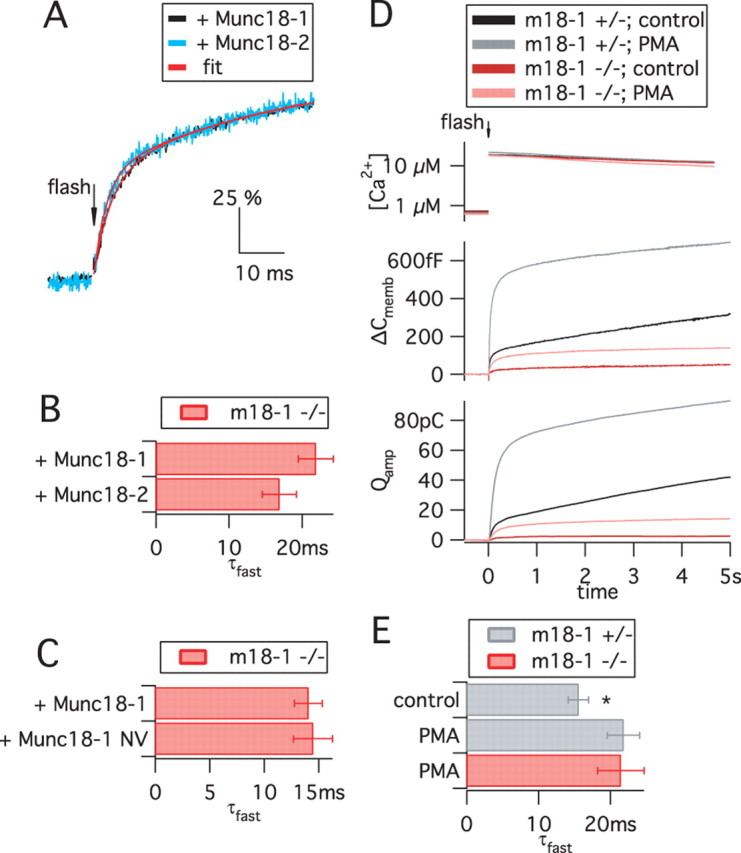
The absence or presence of Munc18 variants does not affect fusion triggering. A–C, Kinetics of the fusion of primed vesicles, triggered by Ca2+ uncaging and assayed by membrane capacitance recordings. Data are from recordings presented in Figures 5 and 6 and supplemental Table 1 (available at www.jneurosci.org as supplemental material). A, The time courses of responses in the presence of Munc18-1 and Munc18-2 (normalized to the burst amplitude, 100%) are similar despite the large difference in the absolute ΔC memb amplitude (refer to Fig. 5 A). A double-exponential function (red lines) was fitted to data points (black and blue traces). B, C, The faster time constant of the exponential fits (τfast, mean ± SEM) was statistically indistinguishable between Munc18-1, Munc18-2, and Munc18-1 NV. For other parameters, refer to supplemental Table 1 (available at www.jneurosci.org as supplemental material). D, PMA treatment increased exocytosis in both munc18-1 −/− and munc18-1 +/− cells by approximately the same factor, showing that the PMA potentiation in embryonic mouse chromaffin cells is not dependent on Munc18-1. Data are means of n = 28 +/− control cells, 31 +/− PMA cells, and 32 −/− PMA cells. E, After PMA treatment, kinetic analysis was possible in munc18-1 −/− cells. Shown is the τfast from +/− and −/− cells. PMA treatment mildly increased the τfast of +/− cells (*p < 0.05), but deletion of Munc18-1 had no effect in the presence of PMA.