
Figure 3.
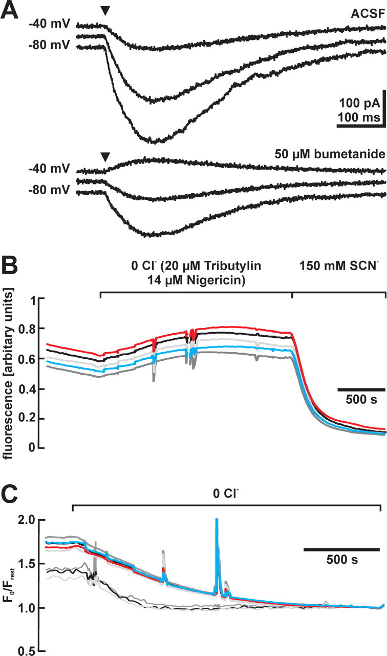
Bumetanide sensitivity of [Cl−]i in CR cells. A, GABAergic currents evoked at a holding potential of −80, −60, and −40 mV before and after the application of bumetanide. Note that EGABA was considerably shifted in a negative direction in the presence of 50 μm bumetanide. B, Representative traces of MEQ fluorescence of five CR cells identified by their morphological appearance in one tangential slice after removal of Cl− and after addition of SCN−. MEQ fluorescence was enhanced in Cl−-free solution and was nearly completely quenched in the presence of SCN−. C, Fluorescence ratio changes of CR cells after removal of Cl− recorded under control conditions (thick lines) and after incubation in 100 μm bumetanide (thin lines). The initial fluorescence ratios, which correspond to resting [Cl−]i, were lower in the bumetanide-incubated cells.