
Figure 4.
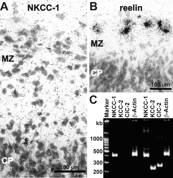
Expression of NKCC1 mRNA in CR cells. A, Bright-field microphotograph of a section from P0 cortex after in situ hybridization using antisense probes for NKCC1 showing the appearance of silver-grains close to the soma of large neurons in the marginal zone (MZ). CP, Cortical plate. B, Bright-field microphotograph of a section from P1 cortex after in situ hybridization using antisense probes for reelin. The intense signals close to the soma of large neurons in the MZ suggest that these neurons are CR cells. C, Ethidiumbromide-stained agarose gel with single-cell multiplex RT-PCR products of cytoplasm harvested from two CR cells. Primers for NKCC1, KCC2, ClC2, and β-actin were used. Note that signals for NKCC1, but also for KCC2 and ClC2, were found in CR cells.