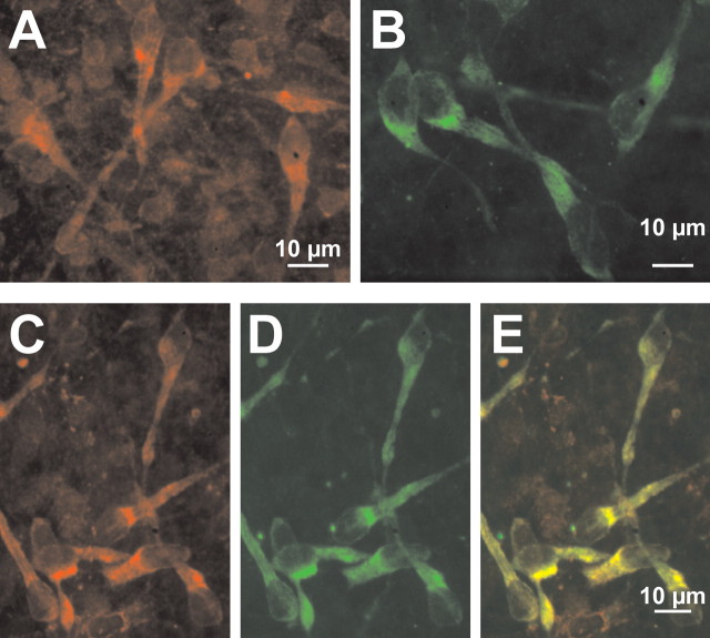
Figure 5.
Immunohistochemical identification of NKCC1 proteins in CR cells. A, Confocal image of NKCC1 immunoreactivity observed with T4 antibody in a tangential slice preparation. Note that large cells with CR cell-like morphology are NKCC1 immunopositive. B, Confocal image of reelin immunoreactivity observed with SP142 antibody in a tangential slice preparation. C–E, Confocal images of tangential slices immunostained simultaneously for NKCC1 (C) and reelin (D). The superimposed image (E) revealed that NKCC1 is expressed in all reelin-immunopositive neurons.

