
Figure 5.
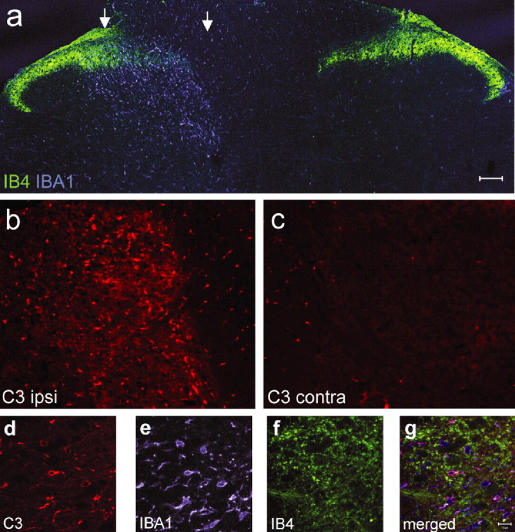
A, Confocal microscopic photomontage of the lumbar dorsal horn of the rat spinal cord 5 d after SNI labeled with the c-fiber central terminal marker IB4 (green) and the microglial marker Iba1 (blue). The region between the arrows has reduced IB4 staining marking the central termination zone of the injured axons. B, C, Complement component C3 immunostaining in the ipsilateral (B) and contralateral (C) dorsal horn 5 d after SNI from the same section acquired using the same microsope settings. D–F, C3 immunoreactivity (5 d SNI; D) colocalizes with Iba1 (microglia; E) but not IB4 (c-fiber; F) staining. G, Overlay of images D–F. Scale bars: A–C, 100 μm; E, F, 10 μm.